Overview
At Vanderbilt University, I worked on CT-based geometry reconstruction and finite-element simulation of the upper airway. The project studied how ansa cervicalis stimulation (ACS) changes airway anatomy in the context of obstructive sleep apnea. I am a joint first author of the resulting paper.
Modeling approach
The study reconstructed anatomy from one adult head-and-neck CT. The workflow combined segmentation, surface smoothing, tetrahedral meshing, and registration of the anatomical components. It used literature-based linear elastic tissue properties.
The model represented ACS as a downward load on the thyroid cartilage and inspiratory loading as a −70 Pa pressure on the airway lumen. Tissue displacement and airway area provided the comparison between loading conditions. Published study.
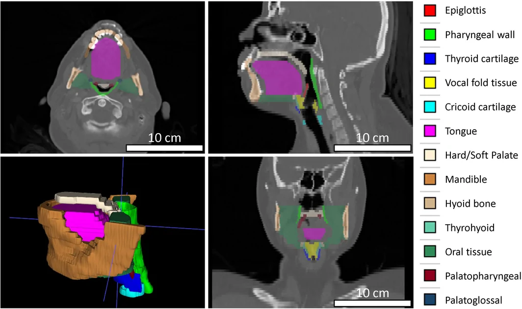
Anatomical segmentation used to construct the finite-element model. Figure 1 from Gupta et al. (2026).
Results
The simulations showed coordinated downward motion of the hyolaryngeal structures and reduced airway narrowing under negative pressure. Epiglottic displacement decreased from 5.0 to 2.2 mm, while retropalatal area increased from 16.98 to 20.43 mm².
These are findings from a single-subject proof-of-concept model. They support investigation of the mechanism rather than a population-level prediction of treatment response. Results and limitations.
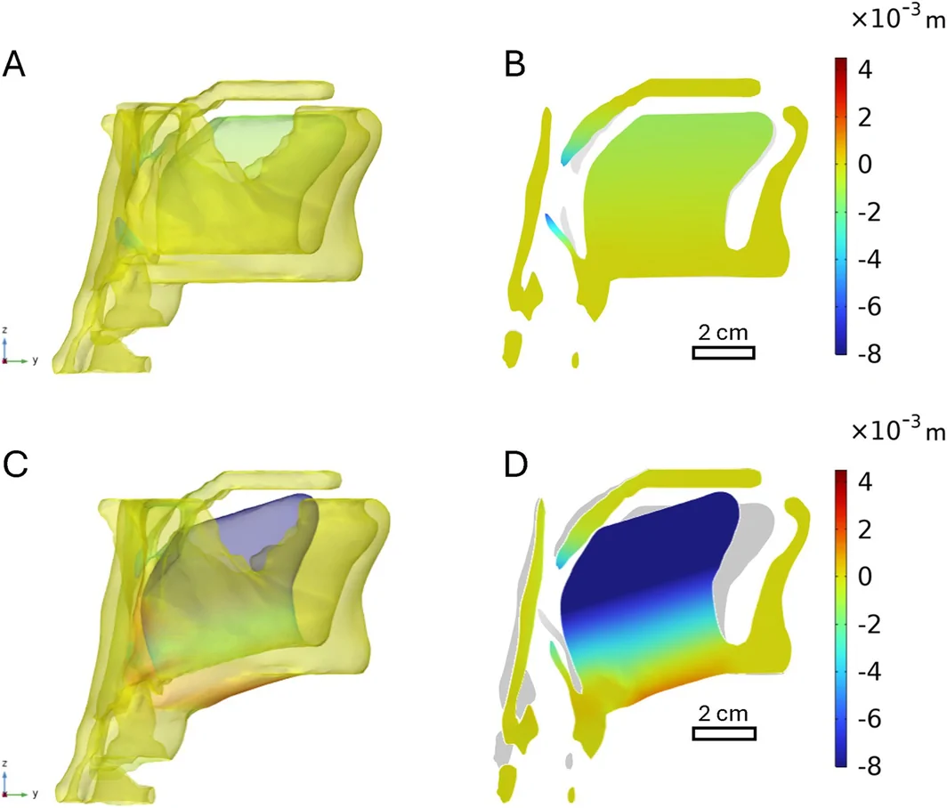
Negative-pressure loading without ACS (A, B) and with ACS (C, D). Figure 6 from Gupta et al. (2026).
Publication
Gupta, M., Li, S., Luo, H., Kent, D. T., and Li, Y. (2026). Finite Element Analysis of Upper Airway in Ansa Cervicalis Stimulation for Obstructive Sleep Apnea. The Laryngoscope, 136(10), 4575–4584. DOI: 10.1002/lary.70603.
Mukund Gupta and Songrui Li contributed equally as joint first authors.